Recently, focusing on the application of organic room-temperature phosphorescence (RTP) in tumor imaging, a collaborative study conducted by the College of Chemistry and Molecular Sciences of Wuhan University (WHU) and the Taikang Institute of Life Sciences was published online on Science Advances. The study was conducted in collaboration with Professor Zhang Yufeng from Wuhan University’s Stomatology Hospital.
The research paper, titled “Lighting up metastasis process before formation of secondary tumor by phosphorescence imaging”, highlights the team’s investigation into the detection of tumor metastasis using organic room-temperature phosphorescence nanoparticles. Chang Kai, a doctoral student from the College of Chemistry and Molecular Sciences, and Xiao Leyi, a doctoral student from the School of Stomatology, are listed as co-first authors. Professor Zhang Yufeng from the School of Stomatology, along with Professors Li Qianqian and Li Zhen from the College of Chemistry and Molecular Sciences, are listed as corresponding authors. Professor Chen Mingzhou from the School of Life Sciences is credited as a contributing author.
According to statistics, tumor metastasis is the leading cause of cancer-related deaths. Despite extensive research on combating cancer metastasis, significant challenges remain in achieving early diagnosis and effective treatment of metastatic cancer. The process of cancer metastasis involves the invasion, intravasation, circulation, extravasation of cancer cells, and the establishment of a premetastatic niche and metastatic niche in distant organs, ultimately leading to the formation of secondary tumors, coexisting with the primary tumor. Currently, the detection of tumor metastasis mainly focuses on the period when secondary tumors have already formed, often resulting in suboptimal treatment outcomes and poor prognosis for patients. Therefore, rapid and accurate identification of the target organs of tumor metastasis at an earlier stage is crucial.
In recent years, organic room-temperature phosphorescence (RTP) biomedical imaging has made significant progress due to its excellent biocompatibility and remarkable background fluorescence suppression ability. RTP has been successfully applied in cell imaging, tumor oxygen imaging, in vivo subcutaneous imaging of mice, in vivo lymphatic imaging, and in vivo subcutaneous tumor imaging. Benefiting from its ability to emit light for a certain period after the excitation source is removed, organic phosphorescence imaging can effectively suppress the background fluorescence of biological samples, thereby exhibiting higher detection sensitivity. Additionally, phosphorescence imaging can achieve high signal-to-noise optical signals at lower power densities by simply controlling the on-off switch of the excitation light source. This technique holds promise for monitoring the process of tumor metastasis and enabling its early diagnosis.
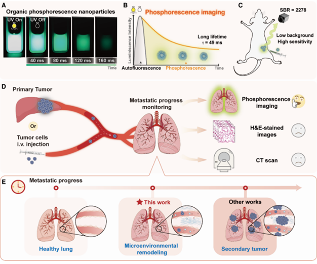
Image 1: Schematic illustration of detecting tumor metastasis process using pure organic room temperature phosphorescence nanoparticles
The research team prepared pure organic phosphorescence nanoparticles based on quinoxaline derivatives, which selectively monitored the lung metastasis of primary tumors. The team implanted H22 liver tumors into the livers of healthy BALB/c mice on day 0, establishing a tumor metastasis model from the liver to the lungs. The degree of lung metastasis was synchronously evaluated every 3 to 4 days using lung phosphorescence imaging, micro-CT, and histopathological examination. Based on the phosphorescence imaging results obtained 3 hours after intravenous injection of the phosphorescence nanoparticles, the maximum phosphorescence radiation intensity in the lungs gradually increased from day 0 to day 27, indicating the gradual accumulation of phosphorescence nanoparticles in the lungs. On day 3, statistical analysis showed a significantly higher maximum phosphorescence radiation intensity of 1.5 ± 0.1 × 10⁵ p sec-1cm-2sr-1 (P=0.0321), demonstrating a significant statistical difference. In the control experiment, the maximum phosphorescence brightness in healthy BALB/c mice injected with phosphorescence nanoparticles remained relatively constant from day 0 to day 27. At this time, no obvious metastatic tumors were observed in the H&E staining analysis. Meanwhile, there were no significant differences in lung volume measured by micro-CT and lung images stained with H&E.
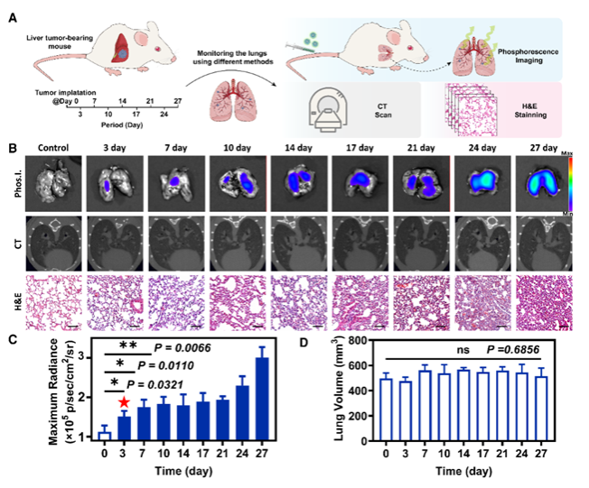
Image 2: Detection of tumor metastasis in a mouse orthotopic liver cancer model using pure organic room-temperature phosphorescent materials, CT, and tissue section H&E staining
To investigate the metastatic status of the lungs, the team conducted further immunohistochemical experiments. The expression of TGF-β, TNF-α, and VEGF was significantly enhanced, with P-values of 0.0002, <0.0001, and 0.0005, respectively, indicating significant changes in the lung microenvironment and the formation of pre-metastatic niches (PMNs). Meanwhile, the expression of BCL-2 slightly increased (P=0.908), suggesting the presence of a small number of circulating tumor cells (CTCs) from liver cancer after the formation of pre-metastatic niches (PMNs). Therefore, the tumor metastasis process detected by phosphorescence imaging corresponds to the formation of metastatic niches in the lungs.
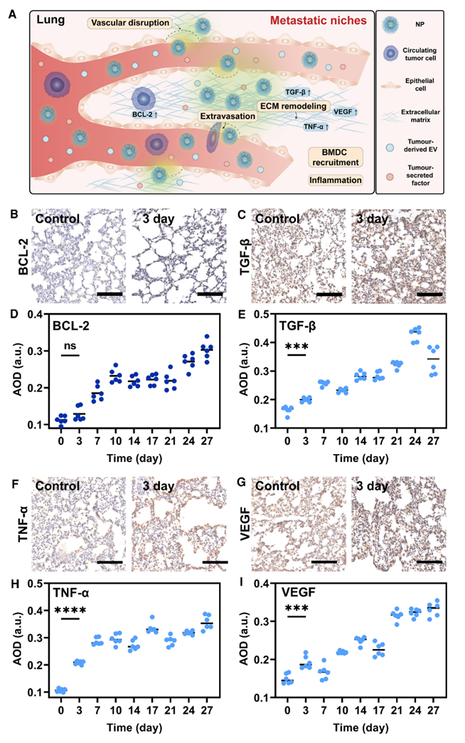
Image 3: Immunohistochemical (IHC) analysis in a mouse orthotopic liver cancer model
In this study, the researchers utilized the long lifetime and high brightness of organic room-temperature phosphorescence nanoparticles to successfully detect the microenvironmental changes prior to the formation of secondary tumors during the process of tumor metastasis. In the orthotopic liver cancer model and the model simulating blood-borne tumor metastasis through intravenous injection of cancer cells, on day 3 after tumor implantation in the liver or intravenous injection of cancer cells, the phosphorescence imaging could detect lung metastasis when only microenvironmental changes were present in the lungs. This method outperforms other imaging techniques such as fluorescence imaging, magnetic resonance imaging (MRI), and PET-CT, which can only detect metastasis at the secondary tumor stage. It provides a sensitive and convenient means for the early detection of tumor metastasis, greatly contributing to preventing the formation and potential worsening of secondary tumors, and providing more opportunities for cancer cure.
This research work was supported by the National Natural Science Foundation of China, the Core Facility of Wuhan University, and the Sharing Platform of Large Equipments in Wuhan University.
Original link: https://www.science.org/doi/10.1126/sciadv.adf6757
Research group website: http://ligroup.whu.edu.cn/
Rewritten by Yu Jiaao
Edited by Shen Le